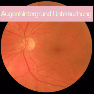
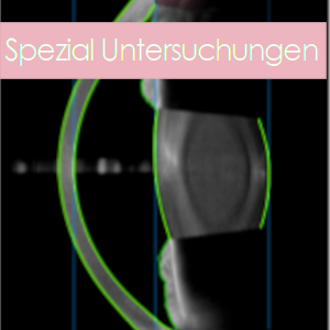
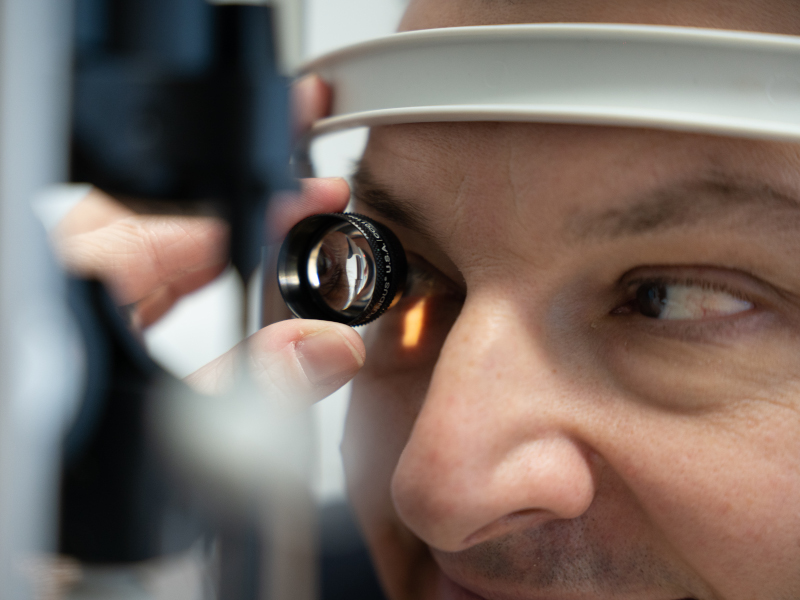
Der Augenhintergrund (Fundus) ist die innere Oberfläche des Augapfels. Mit einer stark vergrößernden Speziallinse können mittels Spaltlampenmikroskop die Netzhaut (Retina) mit dem Sehzentrum (Makula), der Sehnervenkopf (Papille) und die versorgenden Blutgefäße in den hinteren Regionen des Auges beurteilt werden.
Um einen möglichst guten Einblick zu gewinnen, muss die Pupille mithilfe spezieller Augentropfen erweitert werden. Nach der Augenhintergrund Untersuchung können Sie sich geblendet fühlen und das Sehen ist für mehrere Stunden verschwommen. Ein Fahrzeug darf daher nach einer Pupillenerweiterung durch Augentropfen für etwa 2-3 Stunden nicht gelenkt werden.
Insbesondere zur Diagnose und Verlaufskontrolle folgender Erkrankungen ist die Augenhintergrund Untersuchung (Funduskopie) von Bedeutung:
- Makuladegeneration (altersbedingte Erkrankung der Netzhaut im Bereich des Zentrum des schärfsten Sehens, der Makula).
- Veränderungen der Blutgefäße durch Bluthochdruck, Arteriosklerose, Abschätzung des Gefäßstatus sowie des Schlaganfallrisikos, Gefäßverschlüsse der Netzhaut
- Diabetes (diabetische Netzhauterkrankung, aber auch Abschätzung von diabetesbedingten Gefäßschäden in anderen Organsystemen anhand der mikroskopisch vergrößerten Gefäßstrukturen im Auge)
- Erkrankungen des Sehnerven (Grüner Star, Entzündungen, Infarkte)
- Tumore im Augeninneren

